Overview
Background
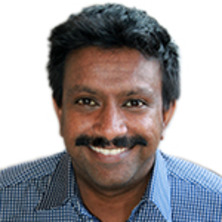
Shakes an imaging expert that leads a strong deep learning, artificial intelligence (AI) focused research team interested in medical image analysis and signal/image processing applied to many areas of science and medicine. He received his Ph.D in Theoretical Physics from Monash University, Melbourne and has been involved in applying machine learning in medical imaging for over a decade.
Shakes’ past work has involved developing shape model-based algorithms for knee, hip and shoulder joint segmentation that is being developed and deployed as a product on the Siemens syngo.via platform. More recent work involves deep learning based algorithms for semantic segmentation and manifold learning of imaging data. Broadly, he is interested in understanding and developing the mathematical basis of imaging, image analysis algorithms and physical systems. He has developed algorithms that utilise exotic mathematical structures such as fractals, turbulence, group theoretic concepts and number theory in the image processing approaches that he has developed.
He is currently a Senior Lecturer and leads a team of 20+ researchers working image analysis and AI research across healthcare and medicine. He currently teaches the computer science courses Theory of Computation and Pattern Recognition and Analysis.
Availability
- Dr Shakes Chandra is:
- Available for supervision
Fields of research
Qualifications
- Doctor of Philosophy, Monash University
Research interests
-
Magnetic Resonance Imaging
Making MRI faster and more affordable through better image reconstruction, processing and analysis.
-
Image Processing
Image reconstruction, segmentation and registration.
-
Deep learning
Dimensionality reduction, machine learning and Artificial Intelligence
-
Fractals and Chaos
Applying fractals and chaos to image processing and computer science.
-
Number Theory
Applying number theory to image processing and computer science.
-
Medical Image Analysis
Medical image segmentation and shape analysis
Works
Search Professor Shakes Chandra’s works on UQ eSpace
2020
Journal Article
Automatic lesion detection, segmentation and characterization via 3D multiscale morphological sifting in breast MRI
Min, Hang, McClymont, Darryl, Chandra, Shekhar S., Crozier, Stuart and Bradley, Andrew P. (2020). Automatic lesion detection, segmentation and characterization via 3D multiscale morphological sifting in breast MRI. Biomedical Physics and Engineering Express, 6 (6) 065027, 065027. doi: 10.1088/2057-1976/abc45c
2020
Journal Article
Fast geometric distortion correction using a deep neural network: implementation for the 1 Tesla MRI-Linac system
Li, Mao, Shan, Shanshan, Chandra, Shekhar S., Liu, Feng and Crozier, Stuart (2020). Fast geometric distortion correction using a deep neural network: implementation for the 1 Tesla MRI-Linac system. Medical Physics, 47 (9) mp.14382, 4303-4315. doi: 10.1002/mp.14382
2020
Conference Publication
Deep simultaneous optimization of sampling and reconstruction for multi-contrast MRI
Liu, Xinwen, Wang, Jing, Tang, Fangfang, Chandra, Shekhar S., Liu, Feng and Crozier, Stuart (2020). Deep simultaneous optimization of sampling and reconstruction for multi-contrast MRI. ISMRM & SMRT Virtual Conference & Exhibition, 2020, Online, 8-14 August 2020.
2020
Journal Article
Simultaneous super-resolution and contrast synthesis of routine clinical magnetic resonance images of the knee for improving automatic segmentation of joint cartilage: data from the Osteoarthritis Initiative
Neubert, Aleš, Bourgeat, Pierrick, Wood, Jason, Engstrom, Craig, Chandra, Shekhar S., Crozier, Stuart and Fripp, Jurgen (2020). Simultaneous super-resolution and contrast synthesis of routine clinical magnetic resonance images of the knee for improving automatic segmentation of joint cartilage: data from the Osteoarthritis Initiative. Medical Physics, 47 (10) mp.14421, 4939-4948. doi: 10.1002/mp.14421
2020
Conference Publication
Fully automatic computer-aided mass detection and segmentation via pseudo-color mammograms and Mask R-CNN
Min, Hang, Wilson, Devin, Huang, Yinhuang, Liu, Siyu, Crozier, Stuart, Bradley, Andrew P. and Chandra, Shekhar S. (2020). Fully automatic computer-aided mass detection and segmentation via pseudo-color mammograms and Mask R-CNN. 17th International Symposium on Biomedical Imaging (ISBI), Iowa City, IA, United States, 3-7 April 2020. Piscataway, NJ, United States: Institute of Electrical and Electronics Engineers (IEEE). doi: 10.1109/isbi45749.2020.9098732
2020
Conference Publication
Fast high dynamic range MRI by Contrast Enhancement Networks
Marques, Matthew, Engstrom, Craig, Fripp, Jurgen, Crozier, Stuart and Chandra, Shekhar S. (2020). Fast high dynamic range MRI by Contrast Enhancement Networks. 2020 IEEE 17th International Symposium on Biomedical Imaging (ISBI), Iowa City, IA, United States, 3-7 April 2020. Piscataway, NJ, United States: Institute of Electrical and Electronics Engineers . doi: 10.1109/isbi45749.2020.9098373
2019
Journal Article
Multi-scale sifting for mammographic mass detection and segmentation
Min, Hang, Chandra, Shekhar S, Crozier, Stuart and Bradley, Andrew P (2019). Multi-scale sifting for mammographic mass detection and segmentation. Biomedical Physics and Engineering Express, 5 (2) 025022, 025022. doi: 10.1088/2057-1976/aafc07
2018
Journal Article
Local contrast-enhanced MR images via high dynamic range processing
Chandra, Shekhar S., Engstrom, Craig, Fripp, Jurgen, Neubert, Ales, Jin, Jin, Walker, Duncan, Salvado, Olivier, Ho, Charles and Crozier, Stuart (2018). Local contrast-enhanced MR images via high dynamic range processing. Magnetic Resonance in Medicine, 80 (3), 1206-1218. doi: 10.1002/mrm.27109
2018
Journal Article
Chaotic Sensing
Chandra, Shekhar S., Ruben, Gary, Jin, Jin, Li, Mingyan, Kingston, Andrew, Svalbe, Imants and Crozier, Stuart (2018). Chaotic Sensing. IEEE Transactions on Image Processing, 27 (12) 8432445, 1-1. doi: 10.1109/TIP.2018.2864918
2018
Journal Article
A lightweight rapid application development framework for biomedical image analysis
Chandra, Shekhar S., Dowling, Jason A., Engstrom, Craig, Xia, Ying, Paproki, Anthony, Neubert, Aleš, Rivest-Hénault, David, Salvado, Olivier, Crozier, Stuart and Fripp, Jurgen (2018). A lightweight rapid application development framework for biomedical image analysis. Computer Methods and Programs in Biomedicine, 164, 193-205. doi: 10.1016/j.cmpb.2018.07.011
2018
Conference Publication
SPIFFY: a simpler image viewer for medical imaging
Sun, Jiayu and Chandra, Shekhar S. (2018). SPIFFY: a simpler image viewer for medical imaging. 4th Information Technology and Mechatronics Engineering Conference (ITOEC2018), Chongqing, China, 14-16 December 2018. Piscataway, NJ, United States: Institute of Electrical and Electronics Engineers (IEEE). doi: 10.1109/ITOEC.2018.8740656
2017
Conference Publication
Multi-scale mass segmentation for mammograms via cascaded random forests
Min, Hang, Chandra, Shekhar S., Dhungel, Neeraj, Crozier, Stuart and Bradley, Andrew P. (2017). Multi-scale mass segmentation for mammograms via cascaded random forests. 14th IEEE International Symposium on Biomedical Imaging, ISBI 2017, Melbourne, VIC, Australia, 18 - 21 April 2017. Piscataway, NJ, United States: Institute of Electrical and Electronics Engineers. doi: 10.1109/ISBI.2017.7950481
2016
Journal Article
Fast automated segmentation of multiple objects via spatially weighted shape learning
Chandra, Shekhar S., Dowling, Jason A., Greer, Peter B., Martin, Jarad, Wratten, Chris, Pichler, Peter, Fripp, Jurgen and Crozier, Stuart (2016). Fast automated segmentation of multiple objects via spatially weighted shape learning. Physics in Medicine and Biology, 61 (22), 8070-8084. doi: 10.1088/0031-9155/61/22/8070
2016
Journal Article
Automatic segmentation of the glenohumeral cartilages from magnetic resonance images
Neubert, A., Yang, Z., Engstrom, C., Xia, Y., Strudwick, M. W., Chandra, S. S., Fripp, J. and Crozier, S. (2016). Automatic segmentation of the glenohumeral cartilages from magnetic resonance images. Medical Physics, 43 (10), 5370-5379. doi: 10.1118/1.4961011
2016
Conference Publication
Incremental shape learning of 3D surfaces of the knee, data from the osteoarthritis initiative
Neubert, Ales, Naser, Ibrahim, Paproki, Anthony, Engstrom, Craig, Fripp, Jurgen, Crozier, Stuart and Chandra, Shekhar S. (2016). Incremental shape learning of 3D surfaces of the knee, data from the osteoarthritis initiative. 2016 IEEE 13th International Symposium on Biomedical Imaging (ISBI), Prague, Czech Republic, 13-16 April, 2016. Piscataway, United States: IEEE Operations Center. doi: 10.1109/ISBI.2016.7493406
2016
Conference Publication
Automated intervertebral disc segmentation using probabilistic shape estimation and active shape models
Neubert, Aleš, Fripp, Jurgen, Chandra, Shekhar S., Engstrom, Craig and Crozier, Stuart (2016). Automated intervertebral disc segmentation using probabilistic shape estimation and active shape models. Third International Workshop and Challenge, CSI 2015, Munich, Germany, 5 October 2015. Switzerland: Springer. doi: 10.1007/978-3-319-41827-8_15
2016
Conference Publication
Finite radial reconstruction for magnetic resonance imaging: a theoretical study
Chandra, Shekhar S., Archchige, Ramitha, Ruben, Gary, Jin, Jin, Li, Mingyan, Kingston, Andrew M., Svalbe, Imants and Crozier, Stuart (2016). Finite radial reconstruction for magnetic resonance imaging: a theoretical study. International Conference on Digital Image Computing: Techniques and Applications, DICTA 2016, Gold Coast, QLD, Australia, 30 November-2 December 2016. Piscataway, NJ, United States: Institute of Electrical and Electronics Engineers. doi: 10.1109/DICTA.2016.7797043
2015
Journal Article
Automatic substitute computed tomography generation and contouring for magnetic resonance imaging (MRI)-alone external beam radiation therapy from standard MRI sequences
Dowling, Jason A., Sun, Jidi, Pichler, Peter, Rivest-Hénault, David, Ghose, Soumya, Richardson, Haylea, Wratten, Chris, Martin, Jarad, Arm, Jameen, Best, Leah, Chandra, Shekhar S., Fripp, Jurgen, Menk, Frederick W. and Greer, Peter B. (2015). Automatic substitute computed tomography generation and contouring for magnetic resonance imaging (MRI)-alone external beam radiation therapy from standard MRI sequences. International Journal of Radiation Oncology Biology Physics, 93 (5), 1144-1153. doi: 10.1016/j.ijrobp.2015.08.045
2015
Journal Article
Automated 3D quantitative assessment and measurement of alpha angles from the femoral head-neck junction using MR imaging
Xia, Ying, Fripp, Jurgen, Chandra, Shekhar S., Walker, Duncan, Crozier, Stuart and Engstrom, Craig M. (2015). Automated 3D quantitative assessment and measurement of alpha angles from the femoral head-neck junction using MR imaging. Physics in Medicine and Biology, 60 (19), 7601-7616. doi: 10.1088/0031-9155/60/19/7601
2015
Journal Article
Automated analysis of hip joint cartilage combining MR T2 and three-dimensional fast-spin-echo images
Chandra, Shekhar S., Surowiec, Rachel, Ho, Charles, Xia, Ying, Engstrom, Craig M., Crozier, Stuart and Fripp, Jurgen (2015). Automated analysis of hip joint cartilage combining MR T2 and three-dimensional fast-spin-echo images. Magnetic Resonance in Medicine, 75 (1), 403-413. doi: 10.1002/mrm.25598
Funding
Current funding
Past funding
Supervision
Availability
- Dr Shakes Chandra is:
- Available for supervision
Looking for a supervisor? Read our advice on how to choose a supervisor.
Available projects
-
Next generation magnetic resonance imaging MRI through vision
Summary: Magnetic resonance imaging (MRI) is crucial for diagnosing diseases within the human body. In this project, we develop new AI methods that leverage human visual perception to make MRI faster and more affordable.
Technologies such as magnetic resonance imaging (MRI) are essential in healthcare for non-invasively seeing inside the human body for disease diagnosis and assessment. However, imaging cost for MRI is so prohibitive that it is seldom used unless there is no other option despite its effectiveness. The cost is largely because MRI is a slow imaging modality compared to other options that do not provide as much information and soft tissue contrast needed to detect diseases such as cancer. Although some progress has been made to improve acquisition speed, all current methods do not make any allowances for the way that human experts read and understand regions of interest. A reduction in scan time will make MRI cheaper and therefore allow the technology to be more readily utilised in the future.
This project aims to create new artificial intelligence (AI) models and unify them with MRI acquisition directly in its measurement domain, helping us explain such models and create acquisitions more akin to human vision that only acquires the areas an operator needs, thereby reducing scan times.
Supervision history
Current supervision
-
Doctor Philosophy
Manifold Learning for Magnetic Resonance Imaging
Principal Advisor
Other advisors: Associate Professor Craig Engstrom, Emeritus Professor Stuart Crozier
-
Doctor Philosophy
Magnetic Resonance Image Processing with Artificial Intelligence
Principal Advisor
Other advisors: Associate Professor Craig Engstrom, Dr Hongfu Sun
-
Doctor Philosophy
Foundational models for visual recognition
Principal Advisor
Other advisors: Associate Professor Craig Engstrom, Emeritus Professor Stuart Crozier
-
Doctor Philosophy
Novel deep learning approaches to understanding human diseases
Principal Advisor
Other advisors: Associate Professor Craig Engstrom, Emeritus Professor Stuart Crozier
-
Doctor Philosophy
AI in the detection and diagnosis of preclinical and early osteoarthritis
Principal Advisor
Other advisors: Associate Professor Craig Engstrom, Dr Kieran O'Brien
-
Doctor Philosophy
Using 3D total body imaging to study the spatial distribution of naevi and melanoma
Associate Advisor
Other advisors: Professor Peter Soyer, Professor Monika Janda
-
Doctor Philosophy
Establishment of a National Anterior Cruciate Ligament (ACL) Registry in Australia
Associate Advisor
Other advisors: Associate Professor Craig Engstrom
-
Doctor Philosophy
Sleep Fragmentation and its Daytime Outcomes in Obstructive Sleep Apnea Patients
Associate Advisor
Other advisors: Professor Juha Toyras, Associate Professor Philip Terrill
-
Doctor Philosophy
Machine learning methods for visualisation and quantification of nephrons with MRI.
Associate Advisor
Other advisors: Dr Nyoman Kurniawan
-
Doctor Philosophy
Neonatal brain MRI of very preterm infants for prediction of neurodevelopmental outcomes
Associate Advisor
Other advisors: Dr Kerstin Pannek
-
Doctor Philosophy
Deep Learning Methods Towards Reliable and Physiology-Aligned Sleep Scoring
Associate Advisor
Other advisors: Professor Juha Toyras, Associate Professor Philip Terrill
Completed supervision
-
2025
Doctor Philosophy
Improving Feature Extraction and Feature Interpretability for Deep Learning in Medical Image Analysis
Principal Advisor
Other advisors: Associate Professor Craig Engstrom, Emeritus Professor Stuart Crozier
-
2025
Doctor Philosophy
Medical Shape Analysis of 3D MRI Segmentation with Deep Learning
Principal Advisor
Other advisors: Emeritus Professor Stuart Crozier
-
2025
Doctor Philosophy
Automated Detection and Classification of Suspicious Naevi in Dermoscopy Images Through Artificial Intelligence
Principal Advisor
Other advisors: Professor Peter Soyer, Professor Monika Janda
-
2024
Doctor Philosophy
Deployable and Interpretable Deep Learning Architectures: Navigating Clinical Challenges in Medical Image Segmentation
Principal Advisor
Other advisors: Associate Professor Craig Engstrom, Emeritus Professor Stuart Crozier
-
2024
Doctor Philosophy
Towards Analysis of Contextual Melanoma Indicators and Identification of Total-Body Ugly Duckling Lesions with Deep Neural Networks
Principal Advisor
Other advisors: Associate Professor Mahsa Baktashmotlagh
-
2024
Doctor Philosophy
Deep Learning Strategies for Enhanced mTBI Diagnosis using Clinical and CT Data
Principal Advisor
Other advisors: Dr Viktor Vegh
-
2024
Doctor Philosophy
Efficient Image Representations for Compressed Sensing MRI
Principal Advisor
Other advisors: Associate Professor Craig Engstrom, Professor Feng Liu, Professor Markus Barth
-
2023
Doctor Philosophy
Towards Efficient Graph Neural Networks for Optimizing Illicit Dark Web Interventions
Principal Advisor
Other advisors: Professor Marius Portmann
-
2022
Doctor Philosophy
Automated Assessment of Cartilage Composition and Cam Morphology using Magnetic Resonance Images of the Hip Joint
Principal Advisor
Other advisors: Associate Professor Craig Engstrom, Emeritus Professor Stuart Crozier
-
2020
Doctor Philosophy
Computer aided lesion detection, segmentation and characterization on mammography and breast MRI
Principal Advisor
Other advisors: Emeritus Professor Stuart Crozier
-
2025
Doctor Philosophy
3D Imaging and Deep Learning for Phenotyping Sorghum at Canopy Scale
Associate Advisor
Other advisors: Professor Scott Chapman
-
2025
Doctor Philosophy
Advanced Deep Learning Approaches for Improving Diagnosis and Prognosis in Brain Disease
Associate Advisor
Other advisors: Professor Fatima Nasrallah
-
2023
Doctor Philosophy
In vitro evaluation of porous PHBV-based scaffolds for tissue regeneration application
Associate Advisor
Other advisors: Associate Professor Mingyuan Lu
-
2022
Doctor Philosophy
Improving deep learning-based fast MRI with pre-acquired image guidance
Associate Advisor
Other advisors: Emeritus Professor Stuart Crozier, Professor Feng Liu
Media
Enquiries
For media enquiries about Dr Shakes Chandra's areas of expertise, story ideas and help finding experts, contact our Media team:
