Overview
Background
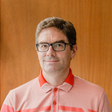
Markus graduated from the Vienna University of Technology in Technical Physics in 1995 and was awarded his Doctorate in 1999 after which he worked as postdoctoral research associate and then Assistant Professor at the Department of Radiodiagnostics, Medical University Vienna (AT). From 2004 he worked as Senior Researcher at the Donders Institute for Brain, Cognition and Behaviour (Radboud University Nijmegen, NL) and at the Erwin L. Hahn Institute for Magnetic Resonance Imaging (University Essen-Duisburg, DE). In 2014 he relocated to the University of Queensland to head the Ultra-high Field Human MR Research program at the Centre for Advanced Imaging and was awarded an ARC Future Fellowship. In 2019 he joined the School of Information Technology and Electrical Engineering as Full Professor Biomedical Engineering working on MR Physics and Medical Imaging. He served as Imaging, Sensing and Biomedical Engineering Discipline lead until 2020 when he took up service roles as Deputy Head of School – Research, Director for the National Imaging Facility – Queensland Node, as well as a member of the ARC College of Experts.
Availability
- Professor Markus Barth is:
- Not available for supervision
- Media expert
Fields of research
Qualifications
- Doctor of Philosophy of Science (Advanced), Technical University Vienna
Research interests
-
Improving MRI
Markus is investigating how MRI can be improved by using new image contrasts by mapping quantitative tissue parameters and by using increased spatial resolution. For example, very small venous vessels and small bleedings in the brain can be visualised using specific contrasts using the MR phase reflecting magnetic susceptibility (SWI and QSM). This information can be used as a very sensitive disease marker in a range of neurodegenerative diseases (traumatic brain injuries, tumours, dementia). He is also developing faster image acquisition methods such as 3D Echo-Planar-Imaging (EPI) that allows reducing the acquisition time by a factor 5-10 compared to standard techniques while keeping the high image fidelity.
-
Understanding brain activity using functional MRI
Blood oxygenation level dependent (BOLD) functional MRI gives a good picture of neural activation and connectivity in the living human brain non-invasively. Markus is particularly interested to identify small functional units of the brain, such as cortical layers and columns, in order to better understand brain function by developing very fast functional MRI techniques with the highest spatial resolution possible. Recently, he also addressed important neuroscientific questions such as memory consolidation during sleep and decoding measured functional signals (brain reading). He also explored the possibilities of simultaneous acquisition of EEG and fMRI to examine the link between electrophysiology and BOLD task activity and large scale brain networks.
Research impacts
Markus has made significant scientific contributions in the fields of Cognitive Neuroscience, Neuroimaging, and MR methods at (ultra-)high field and key contributions to MRI scanner software packages, which are used in MR labs worldwide. Markus’ main interest is to improve our understanding of brain function and disfunction in cognition, neurodegenerative diseases, and cancer by developing new medical imaging techniques. With a focus on human neuroimaging using magnetic resonance imaging (MRI) at high and ultra-high magnetic field strength, he achieved fast, high resolution mapping of magnetic susceptibility related anatomical and functional information in vivo, including characterisation of blood oxygenation, iron storage in tissue, haemorrhage and calcifications. Recent achievements include the development of accurate detection of layer specific functional activation in the human brain, decoding of brain activity and ultra-fast MRI. His research interests are in the fields of MR method development including applications in neuroimaging and neurological diseases including dementia, motor neurone disease, and cancer.
Works
Search Professor Markus Barth’s works on UQ eSpace
2010
Journal Article
A population-specific symmetric phase model to automatically analyze susceptibility-weighted imaging (SWI) phase shifts and phase symmetry in the human brain
Grabner, Gunther, Haubenberger, Dietrich, Rath, Jakob, Beisteiner, Roland, Auff, Eduard, Trattnig, Siegfried and Barth, Markus (2010). A population-specific symmetric phase model to automatically analyze susceptibility-weighted imaging (SWI) phase shifts and phase symmetry in the human brain. Journal of Magnetic Resonance Imaging, 31 (1), 215-220. doi: 10.1002/jmri.22013
2010
Journal Article
T2-weighted 3D fMRI using S2-SSFP at 7 tesla
Barth, Markus, Meyer, Heiko, Kannengiesser, Stephan A. R., Polimeni, Jonathan R., Wald, Lawrence L. and Norris, David G. (2010). T2-weighted 3D fMRI using S2-SSFP at 7 tesla. Magnetic Resonance in Medicine, 63 (4), 1015-1020. doi: 10.1002/mrm.22283
2010
Journal Article
Susceptibility weighted magnetic resonance imaging of cerebral cavernous malformations: prospects, drawbacks, and first experience at ultra-high field strength (7-Tesla) magnetic resonance imaging.
Dammann,Philipp, Barth, Markus, Zhu, Yuan, Maderwald, Stefan, Schlamann, Marc, Ladd, Mark E. and Sure, Ulrich (2010). Susceptibility weighted magnetic resonance imaging of cerebral cavernous malformations: prospects, drawbacks, and first experience at ultra-high field strength (7-Tesla) magnetic resonance imaging.. Neurosurgical focus, 29 (3), 1-7. doi: 10.3171/2010.6.FOCUS10130
2009
Journal Article
Phase unwrapping of MR images using ΦUN - A fast and robust region growing algorithm
Witoszynskyj, Stephan, Rauscher, Alexander, Reichenbach, Jürgen R. and Barth, Markus (2009). Phase unwrapping of MR images using ΦUN - A fast and robust region growing algorithm. Medical Image Analysis, 13 (2), 257-268. doi: 10.1016/j.media.2008.10.004
2008
Journal Article
MR venography of the human brain using susceptibility weighted imaging at very high field strength
Koopmans, Peter J., Manniesing, Rashindra, Niessen, Wiro J., Viergever, Max A. and Barth, Markus (2008). MR venography of the human brain using susceptibility weighted imaging at very high field strength. Magnetic Resonance Materials in Physics, Biology and Medicine, 21 (1-2), 149-158. doi: 10.1007/s10334-007-0101-3
2008
Journal Article
T1 mapping of the entire lung parenchyma: Influence of respiratory phase and correlation to lung function test results in patients with diffuse lung disease
Stadler, Alfred, Jakob, Peter M., Griswold, Mark, Stiebellehner, Leopold, Barth, Markus and Bankier, Alexander A. (2008). T1 mapping of the entire lung parenchyma: Influence of respiratory phase and correlation to lung function test results in patients with diffuse lung disease. Magnetic Resonance in Medicine, 59 (1), 96-101. doi: 10.1002/mrm.21446
2008
Journal Article
Improved elimination of phase effects from background field inhomogeneities for susceptibility weighted imaging at high magnetic field strengths
Rauscher, Alexander, Barth, Markus, Herrmann, Karl-Heinz, Witoszynskyj, Stephan, Deistung, Andreas and Reichenbach, Jüergen R. (2008). Improved elimination of phase effects from background field inhomogeneities for susceptibility weighted imaging at high magnetic field strengths. Magnetic Resonance Imaging, 26 (8), 1145-1151. doi: 10.1016/j.mri.2008.01.029
2007
Journal Article
Very high-resolution three-dimensional functional MRI of the human visual cortex with elimination of large venous vessels
Barth, M. and Norris, D. G. (2007). Very high-resolution three-dimensional functional MRI of the human visual cortex with elimination of large venous vessels. NMR in Biomedicine, 20 (5), 477-484. doi: 10.1002/nbm.1158
2007
Journal Article
Contrast-to-noise ratio (CNR) as a quality parameter in fMRI
Geissler, Alexander, Gartus, Andreas, Foki, Thomas, Tahamtan, Amir Reza, Beisteiner, Roland and Barth, Markus (2007). Contrast-to-noise ratio (CNR) as a quality parameter in fMRI. Journal of Magnetic Resonance Imaging, 25 (6), 1263-1270. doi: 10.1002/jmri.20935
2007
Journal Article
Comparison of fMRI coregistration results between human experts and software solutions in patients and healthy subjects
Gartus, Andreas, Geissler, Alexander, Foki, Thomas, Tahamtan, Amir Reza, Pahs, Gerald, Barth, Markus, Pinker, Katja, Trattnig, Siegfried and Beisteiner, Roland (2007). Comparison of fMRI coregistration results between human experts and software solutions in patients and healthy subjects. European Radiology, 17 (6), 1634-1643. doi: 10.1007/s00330-006-0459-z
2006
Journal Article
Contrast-enhanced, high-resolution, susceptibility-weighted magnetic resonance imaging of the brain: dose-dependent optimization at 3 Tesla and 1.5 Tesla in healthy volunteers
Noebauer-Huhmann, Iris-Melanie, Pinker, Katja, Barth, Markus, Mlynarik, Vladimir, Ba-Ssalamah, Ahmed, Saringer, Walter F., Weber, Michael, Benesch, Thomas, Witoszynskyj, Stephan, Rauscher, Alexander, Reichenbach, Juergen and Trattnig, Siegfried (2006). Contrast-enhanced, high-resolution, susceptibility-weighted magnetic resonance imaging of the brain: dose-dependent optimization at 3 Tesla and 1.5 Tesla in healthy volunteers. Investigative Radiology, 41 (3), 249-255. doi: 10.1097/01.rli.0000188360.24222.5e
2006
Journal Article
Contrast Enhanced Susceptibility Weighted Imaging (CE-SWI) of the Mouse Brain Using Ultrasmall Superparamagnetic Ironoxide Particles (USPIO)
Hamans, Bob C., Barth, Markus, Leenders, William P. and Heerschap, Arend (2006). Contrast Enhanced Susceptibility Weighted Imaging (CE-SWI) of the Mouse Brain Using Ultrasmall Superparamagnetic Ironoxide Particles (USPIO). Zeitschrift Fur Medizinische Physik, 16 (4), 269-274. doi: 10.1078/0939-3889-00325
2005
Journal Article
Evaluation of preoperative high magnetic field motor functional MRI (3 Tesla) in glioma patients by navigated electrocortical stimulation and postoperative outcome
Roessler, K., Donat, M., Lanzenberger, R., Novak, K., Geissler, A., Gartus, A., Tahamtan, A. R., Milakara, D., Czech, T., Barth, M., Knosp, E. and Beisteiner, R. (2005). Evaluation of preoperative high magnetic field motor functional MRI (3 Tesla) in glioma patients by navigated electrocortical stimulation and postoperative outcome. Journal of Neurology, Neurosurgery and Psychiatry, 76 (8), 1152-1157. doi: 10.1136/jnnp.2004.050286
2005
Journal Article
Nonnvasive assessment of vascular architecture and function during modulated blood oxygenation using susceptibility weighted magnetic resonance imaging
Rauscher, Alexander, Sedlacik, Jan, Barth, Markus, Haacke, E. Mark and Reichenbach, Juergen R. (2005). Nonnvasive assessment of vascular architecture and function during modulated blood oxygenation using susceptibility weighted magnetic resonance imaging. Magnetic Resonance in Medicine, 54 (1), 87-95. doi: 10.1002/mrm.20520
2005
Journal Article
T1 mapping of the entire lung parenchyma: influence of the respiratory phase in healthy individuals
Stadler, Alfred, Jakob, Peter M., Griswold, Mark, Barth, Markus and Bankier, Alexander A. (2005). T1 mapping of the entire lung parenchyma: influence of the respiratory phase in healthy individuals. Journal of Magnetic Resonance Imaging, 21 (6), 759-764. doi: 10.1002/jmri.20319
2005
Journal Article
Magnetic susceptibility-weighted MR phase imaging of the human brain
Rauscher, A, Sedlacik, J, Barth, M, Mentzel, HJ and Reichenbach, JR (2005). Magnetic susceptibility-weighted MR phase imaging of the human brain. American Journal of Neuroradiology, 26 (4), 736-742.
2005
Journal Article
FMRI reveals functional cortex in a case of inconclusive Wada testing
Lanzenberger, Rupert, Wiest, Gerald, Geissler, Alexander, Barth, Markus, Ringl, Helmut, Wober, Christian, Gartus, Andreas, Baumgartner, Christoph and Beisteiner, Roland (2005). FMRI reveals functional cortex in a case of inconclusive Wada testing. Clinical Neurology and Neurosurgery, 107 (2), 147-151. doi: 10.1016/j.clineuro.2004.06.006
2005
Journal Article
Influence of fMRI smoothing procedures on replicability of fine scale motor localization
Geissler, Alexander, Lanzenberger, Rupert, Barth, Markus, Tahamtan, Amir Reza, Milakara, Denny, Gartus, Andreas and Beisteiner, Roland (2005). Influence of fMRI smoothing procedures on replicability of fine scale motor localization. NeuroImage, 24 (2), 323-331. doi: 10.1016/j.neuroimage.2004.08.042
2005
Book Chapter
Probleme und Lösungsmöglichkeiten bei Patientenuntersuchungen mit funktioneller Magnetresonanztomographie (fMRT)
Beisteiner, Roland and Barth, Markus (2005). Probleme und Lösungsmöglichkeiten bei Patientenuntersuchungen mit funktioneller Magnetresonanztomographie (fMRT). Funktionelle Bildgebung in Psychiatrie und Psychotherapie – Methodische Grundlagen und Klinische Anwendungen. (pp. 74-88) edited by Henrik Walter. Stuttgart: Schattauer Verlag.
2004
Journal Article
Robust field map generation using a triple-echo acquisition
Windischberger, Christian, Robinson, Simon, Rauscher, Alexander, Barth, Markus and Moser, Ewald (2004). Robust field map generation using a triple-echo acquisition. Journal of Magnetic Resonance Imaging, 20 (4), 730-734. doi: 10.1002/jmri.20158
Funding
Current funding
Supervision
Availability
- Professor Markus Barth is:
- Not available for supervision
Supervision history
Current supervision
-
Doctor Philosophy
Evaluating Magnetic Resonance Spectroscopy and Spectroscopic Imaging Techniques in Motor Neuron Disease at 3T and 7T
Principal Advisor
Other advisors: Dr Kieran O'Brien, Dr Thomas Shaw
-
Doctor Philosophy
Additive manufacturing in the patient specific optimisation of intracavitary brachytherapy
Associate Advisor
Other advisors: Dr Scott Crowe
-
Doctor Philosophy
Improving vascular MRI with deep learning.
Associate Advisor
Other advisors: Dr Fernanda Lenita Ribeiro, Dr Saskia Bollmann
Completed supervision
-
2025
Doctor Philosophy
Towards QSM validation: reference measurement, doping materials, and anthropomorphic phantoms
Principal Advisor
Other advisors: Dr Kieran O'Brien, Dr Monique Tourell
-
2025
Doctor Philosophy
Automation of Daily MRI Quality Assurance and Standardisation of Preclinical PET/CT Reconstruction Protocols Using Phantom-Based Evaluation
Principal Advisor
Other advisors: Dr Viktor Vegh, Dr Monique Tourell
-
2025
Doctor Philosophy
Neural Network-Enhanced Multimodal Brain Electrical Source Imaging and Applications
Principal Advisor
Other advisors: Dr Steffen Bollmann
-
2023
Doctor Philosophy
Improving functional MRI through Modelling and Imaging Microvascular Dynamics
Principal Advisor
Other advisors: Dr Saskia Bollmann
-
2022
Doctor Philosophy
Modelling the Depth-dependent Functional Responses in Human Primary Visual and Motor Cortices
Principal Advisor
Other advisors: Dr Saskia Bollmann
-
2021
Doctor Philosophy
Sequence Development to Improve Image Quality for T2- and Diffusion Weighted Imaging at 7T
Principal Advisor
Other advisors: Dr Kieran O'Brien, Dr Steffen Bollmann
-
2020
Doctor Philosophy
MR signal modelling approaches to characterise tissue microstructure in in-vivo human brain
Principal Advisor
Other advisors: Dr Viktor Vegh, Dr Steffen Bollmann
-
2018
Doctor Philosophy
Evaluating Acquisition Techniques for Functional Magnetic Resonance Imaging at Ultra-High Field
Principal Advisor
-
2019
Doctor Philosophy
Radiofrequency safety and shimming near metal hip prostheses at high and ultra-high field MRI
Joint Principal Advisor
Other advisors: Professor Feng Liu, Dr Kieran O'Brien, Emeritus Professor Stuart Crozier
-
2024
Doctor Philosophy
Efficient Image Representations for Compressed Sensing MRI
Associate Advisor
Other advisors: Associate Professor Craig Engstrom, Professor Feng Liu, Dr Shakes Chandra
-
2024
Doctor Philosophy
Parallel Transmission for Advanced MRI Techniques at Ultra-High Field
Associate Advisor
-
2023
Master Philosophy
Solving Quantitative Susceptibility Mapping using Deep Learning
Associate Advisor
Other advisors: Dr Steffen Bollmann
-
2023
Doctor Philosophy
Automated Quantitative Susceptibility Mapping for Clinical Applications
Associate Advisor
Other advisors: Dr Kieran O'Brien, Dr Monique Tourell, Dr Steffen Bollmann
-
2021
Doctor Philosophy
Computational in vivo Tissue Characterisation for Multi-Contrast High-Resolution Magnetic Resonance Imaging Data
Associate Advisor
Other advisors: Dr Steffen Bollmann
-
2019
Doctor Philosophy
Measuring tissue variations in the human brain using quantitative MRI
Associate Advisor
Other advisors: Dr Viktor Vegh
-
2018
Doctor Philosophy
Neural correlates of visual function in agenesis of the corpus callosum
Associate Advisor
Other advisors: Professor Jason Mattingley
Media
Enquiries
Contact Professor Markus Barth directly for media enquiries about:
- Biomedical engineering
- Biomedical Imaging
- Brain imaging
- Diffusion imaging
- fMRI
- Image analysis
- Image reconstruction
- Imaging Processing
- Magnetic Resonance Imaging
- MR Imaging Techniques
- MRI
- Neuroimaging
Need help?
For help with finding experts, story ideas and media enquiries, contact our Media team:
